We have a new app!
Take the Access library with you wherever you go—easy access to books, videos, images, podcasts, personalized features, and more.
Download the Access App here: iOS and Android . Learn more here!
- Remote Access
- Save figures into PowerPoint
- Download tables as PDFs


2: Acute Otitis Media
Aimee Dassner; Jennifer E. Girotto
- Download Chapter PDF
Disclaimer: These citations have been automatically generated based on the information we have and it may not be 100% accurate. Please consult the latest official manual style if you have any questions regarding the format accuracy.
Download citation file:
- Search Book
Jump to a Section
Patient presentation.
- Full Chapter
- Supplementary Content
Chief Complaint
“Increased irritability and right ear pain.”
History of Present Illness
JL is a 22-month-old female who presents to her primary care provider (PCP) with a 2-day history of rhinorrhea and a 1-day history of increased irritability, fever (to 101.5°F per Mom), and right-ear tugging. Mom denies that JL has had any nausea, vomiting, or diarrhea.
Past Medical History
Full-term birth via spontaneous vaginal delivery. Hospitalized at 9 months of age for respiratory syncytial virus–associated bronchiolitis. Two episodes of acute otitis media (AOM), with last episode about 6 months earlier.
Surgical History
Social history.
Lives with mother, father, and her 5-year-old brother who attends kindergarten. JL attends daycare 2 d/wk, and stays at home with maternal grandmother 3 d/wk.
No known drug allergies
Immunizations
Home medications.
Vitamin D drops 600 IU/d
Physical Examination
Vital signs (while crying).
Temp 100.7°F, P 140 bpm, RR 35, BP 100/57 mm Hg, Ht 81 cm, Wt 23.7 kg
Fussy, but consolable by Mom; well-appearing
Normocephalic, atraumatic, moist mucous membranes, normal conjunctiva, clear rhinorrhea, moderate bulging and erythema of right tympanic membrane with middle-ear effusion
Good air movement throughout, clear breath sounds bilaterally
Cardiovascular
Normal rate and rhythm, no murmur, rub or gallop
Soft, non-distended, non-tender, active bowel sounds
Genitourinary
Normal female genitalia, no dysuria or hematuria
Alert and appropriate for age
Extremities
1. Which of the following clinical criteria is not part of the diagnostic evaluation or staging of acute otitis media (AOM) for this patient?
A. Rhinorrhea
D. Contour of the tympanic membrane
Sign in or create a free Access profile below to access even more exclusive content.
With an Access profile, you can save and manage favorites from your personal dashboard, complete case quizzes, review Q&A, and take these feature on the go with our Access app.
Pop-up div Successfully Displayed
This div only appears when the trigger link is hovered over. Otherwise it is hidden from view.
Please Wait

- My presentations
Auth with social network:
Download presentation
We think you have liked this presentation. If you wish to download it, please recommend it to your friends in any social system. Share buttons are a little bit lower. Thank you!
Presentation is loading. Please wait.
ACUTE AND CHRONIC OTITIS MEDIA
Published by Pauli Kivelä Modified over 5 years ago
Similar presentations
Presentation on theme: "ACUTE AND CHRONIC OTITIS MEDIA"— Presentation transcript:
By : wala’ mosa Presented to: Dr. Ayham Abu Lila.

Otology Dave Pothier St Mary’s 2003.

DRUGS DO NOT DO DRUGS !!! Hearing disorders in children/ Hala AlOmari.

Hearing disorders of the middle ear

به نام خدا.

Chronic otitis media Chunfu Dai M.D & Ph. D Otolaryngology Department

Otitis Media.

Department of Otorhinolaryngology

Daekeun Joo Resident Lecture Series 11/18/09

CAUSES OF HEARING IMPAIRMENT

Otitis Media and Eustachian Tube Dysfunction

The complications of acute and chronic otitis media

Cholesteatoma and chronic suppurative otitis media

Objectives Upon completion of the lecture, students should be able to: Define middle ear infection Know the classification of otitis media (OM).

Definitions Middle ear is the area between the tympanic membrane and the inner ear including the Eustachian tube. Otitis media (OM) is inflammation.

Treatment Antibiotics Antibiotics Surgery Surgery Myringotomy and suction Myringotomy and suction Mastoidectomy (if infection has spread to mastoid region)

Otitis media.

Babak Saedi Imam Khomeini Hospital

بسم الله الرحمن الرحيم.

King Abdulaziz University Hospital

About project
© 2024 SlidePlayer.com Inc. All rights reserved.
An official website of the United States government
The .gov means it's official. Federal government websites often end in .gov or .mil. Before sharing sensitive information, make sure you're on a federal government site.
The site is secure. The https:// ensures that you are connecting to the official website and that any information you provide is encrypted and transmitted securely.
- Publications
- Account settings
- Browse Titles
NCBI Bookshelf. A service of the National Library of Medicine, National Institutes of Health.
StatPearls [Internet]. Treasure Island (FL): StatPearls Publishing; 2024 Jan-.

StatPearls [Internet].
Acute otitis media.
Amina Danishyar ; John V. Ashurst .
Affiliations
Last Update: April 15, 2023 .
- Continuing Education Activity
Acute otitis media (AOM) is defined as an infection of the middle ear and is the second most common pediatric diagnosis in the emergency department following upper respiratory infections. Although acute otitis media can occur at any age, it is most commonly seen between the ages of 6 to 24 months. Approximately 80% of all children will experience a case of otitis media during their lifetime, and between 80% and 90% of all children will have otitis media with an effusion before school age. This activity reviews the etiology, epidemiology, evaluation, and management of acute otitis media and highlights the role of the interprofessional team in managing this condition.
- Describe a patient presentation consistent with acute otitis media and the subsequent evaluation that should be performed.
- Explain when imaging studies should be done for a patient with acute otitis media.
- Outline the treatment strategy for otitis media.
- Employ an interprofessional team approach when caring for patients with acute otitis media.
- Introduction
Acute otitis media is defined as an infection of the middle ear space. It is a spectrum of diseases that includes acute otitis media (AOM), chronic suppurative otitis media (CSOM), and otitis media with effusion (OME). Acute otitis media is the second most common pediatric diagnosis in the emergency department, following upper respiratory infections. Although otitis media can occur at any age, it is most commonly seen between the ages of 6 to 24 months. [1]
Infection of the middle ear can be viral, bacterial, or coinfection. The most common bacterial organisms causing otitis media are Streptococcus pneumoniae , followed by non-typeable Haemophilus influenzae (NTHi) and Moraxella catarrhalis . Following the introduction of the conjugate pneumococcal vaccines, the pneumococcal organisms have evolved to non-vaccine serotypes. The most common viral pathogens of otitis media include the respiratory syncytial virus (RSV), coronaviruses, influenza viruses, adenoviruses, human metapneumovirus, and picornaviruses. [2] [3] [4]
Otitis media is diagnosed clinically via objective findings on physical exam (otoscopy) combined with the patient's history and presenting signs and symptoms. Several diagnostic tools are available such as a pneumatic otoscope, tympanometry, and acoustic reflectometry, to aid in the diagnosis of otitis media. Pneumatic otoscopy is the most reliable and has a higher sensitivity and specificity as compared to plain otoscopy, though tympanometry and other modalities can facilitate diagnosis if pneumatic otoscopy is unavailable.
Treatment of otitis media with antibiotics is controversial and directly related to the subtype of otitis media in question. Without proper treatment, suppurative fluid from the middle ear can extend to the adjacent anatomical locations and result in complications such as tympanic membrane (TM) perforation, mastoiditis, labyrinthitis, petrositis, meningitis, brain abscess, hearing loss, lateral and cavernous sinus thrombosis, and others. [5] This has led to the development of specific guidelines for the treatment of OM. In the United States, the mainstay of treatment for an established diagnosis of AOM is high-dose amoxicillin, and this has been found to be most effective in children under two years of age. Treatment in countries like the Netherlands is initially watchful waiting, and if unresolved, antibiotics are warranted [6] . However, the concept of watchful waiting has not gained full acceptance in the United States and other countries due to the risk of prolonged middle ear fluid and its effect on hearing and speech, as well as the risks of complications discussed earlier. Analgesics such as non-steroidal anti-inflammatory medications such as ibuprofen can be used alone or in combination to achieve effective pain control in patients with otitis media.
Otitis media is a multifactorial disease. Infectious, allergic, and environmental factors contribute to otitis media. [7] [8] [9] [10] [11] [12]
These causes and risk factors include:
- Decreased immunity due to human immunodeficiency virus (HIV), diabetes, and other immuno-deficiencies
- Genetic predisposition
- Mucins that include abnormalities of this gene expression, especially upregulation of MUC5B
- Anatomic abnormalities of the palate and tensor veli palatini
- Ciliary dysfunction
- Cochlear implants
- Vitamin A deficiency
- Bacterial pathogens, Streptococcus pneumoniae , Haemophilus influenza, and Moraxella (Branhamella) catarrhalis are responsible for more than 95%
- Viral pathogens such as respiratory syncytial virus, influenza virus, parainfluenza virus, rhinovirus, and adenovirus
- Lack of breastfeeding
- Passive smoke exposure
- Daycare attendance
- Lower socioeconomic status
- Family history of recurrent AOM in parents or siblings
- Epidemiology
Otitis media is a global problem and is found to be slightly more common in males than in females. The specific number of cases per year is difficult to determine due to the lack of reporting and different incidences across many different geographical regions. The peak incidence of otitis media occurs between six and twelve months of life and declines after age five. Approximately 80% of all children will experience a case of otitis media during their lifetime, and between 80% and 90% of all children will experience otitis media with an effusion before school age. Otitis media is less common in adults than in children, though it is more common in specific sub-populations such as those with a childhood history of recurrent OM, cleft palate, immunodeficiency or immunocompromised status, and others. [13] [14]
- Pathophysiology
Otitis media begins as an inflammatory process following a viral upper respiratory tract infection involving the mucosa of the nose, nasopharynx, middle ear mucosa, and Eustachian tubes. Due to the constricted anatomical space of the middle ear, the edema caused by the inflammatory process obstructs the narrowest part of the Eustachian tube leading to a decrease in ventilation. This leads to a cascade of events resulting in an increase in negative pressure in the middle ear, increasing exudate from the inflamed mucosa, and buildup of mucosal secretions, which allows for the colonization of bacterial and viral organisms in the middle ear. The growth of these microbes in the middle ear then leads to suppuration and, eventually, frank purulence in the middle ear space. This is demonstrated clinically by a bulging or erythematous tympanic membrane and purulent middle ear fluid. This must be differentiated from chronic serous otitis media (CSOM), which presents with thick, amber-colored fluid in the middle ear space and a retracted tympanic membrane on otoscopic examination. Both will yield decreased TM mobility on tympanometry or pneumatic otoscopy.
Several risk factors can predispose children to develop acute otitis media. The most common risk factor is a preceding upper respiratory tract infection. Other risk factors include male gender, adenoid hypertrophy (obstructing), allergy, daycare attendance, environmental smoke exposure, pacifier use, immunodeficiency, gastroesophageal reflux, parental history of recurrent childhood OM, and other genetic predispositions. [15] [16] [17]
- Histopathology
Histopathology varies according to disease severity. Acute purulent otitis media (APOM) is characterized by edema and hyperemia of the subepithelial space, which is followed by the infiltration of polymorphonuclear (PMN) leukocytes. As the inflammatory process progresses, there is mucosal metaplasia and the formation of granulation tissue. After five days, the epithelium changes from flat cuboidal to pseudostratified columnar with the presence of goblet cells.
In serous acute otitis media (SAOM), inflammation of the middle ear and the eustachian tube has been identified as the major precipitating factor. Venous or lymphatic stasis in the nasopharynx or the eustachian tube plays a vital role in the pathogenesis of AOM. Inflammatory cytokines attract plasma cells, leukocytes, and macrophages to the site of inflammation. The epithelium changes to pseudostratified, columnar, or cuboidal. Hyperplasia of basal cells results in an increased number of goblet cells in the new epithelium. [18]
In practice, biopsy for histology is not performed for OM outside of research settings.
- History and Physical
Although one of the best indicators for otitis media is otalgia, many children with otitis media can present with non-specific signs and symptoms, which can make the diagnosis challenging. These symptoms include pulling or tugging at the ears, irritability, headache, disturbed or restless sleep, poor feeding, anorexia, vomiting, or diarrhea. Approximately two-thirds of the patients present with fever, which is typically low-grade.
The diagnosis of otitis media is primarily based on clinical findings combined with supporting signs and symptoms as described above. No lab test or imaging is needed. According to guidelines set forth by the American Academy of Pediatrics, evidence of moderate to severe bulging of the tympanic membrane or new onset of otorrhea not caused by otitis externa or mild tympanic membrane (TM) bulging with recent onset of ear pain or erythema is required for the diagnosis of acute otitis media. These criteria are intended only to aid primary care clinicians in the diagnosis and proper clinical decision-making but not to replace clinical judgment. [19]
Otoscopic examination should be the first and most convenient way of examining the ear and will yield the diagnosis to the experienced eye. In AOM, the TM may be erythematous or normal, and there may be fluid in the middle ear space. In suppurative OM, there will be obvious purulent fluid visible and a bulging TM. The external ear canal (EAC) may be somewhat edematous, though significant edema should alert the clinician to suspect otitis externa (outer ear infection, AOE), which may be treated differently. In the presence of EAC edema, it is paramount to visualize the TM to ensure it is intact. If there is an intact TM and a painful, erythematous EAC, ototopical drops should be added to treat AOE. This can exist in conjunction with AOM or independent of it, so visualization of the middle ear is paramount. If there is a perforation of the TM, then the EAC edema can be assumed to be reactive, and ototopical medication should be used, but an agent approved for use in the middle ear, such as ofloxacin, must be used, as other agents can be ototoxic. [20] [21] [22]
The diagnosis of otitis media should always begin with a physical exam and the use of an otoscope, ideally a pneumatic otoscope. [23] [24]
Laboratory Studies
Laboratory evaluation is rarely necessary. A full sepsis workup in infants younger than 12 weeks with fever and no obvious source other than associated acute otitis media may be necessary. Laboratory studies may be needed to confirm or exclude possible related systemic or congenital diseases.
Imaging Studies
Imaging studies are not indicated unless intra-temporal or intracranial complications are a concern. [25] [26]
- When an otitis media complication is suspected, computed tomography of the temporal bones may identify mastoiditis, epidural abscess, sigmoid sinus thrombophlebitis, meningitis, brain abscess, subdural abscess, ossicular disease, and cholesteatoma.
- Magnetic resonance imaging may identify fluid collections, especially in the middle ear collections.
Tympanocentesis
Tympanocentesis may be used to determine the presence of middle ear fluid, followed by culture to identify pathogens.
Tympanocentesis can improve diagnostic accuracy and guide treatment decisions but is reserved for extreme or refractory cases. [27] [28]
Other Tests
Tympanometry and acoustic reflectometry may also be used to evaluate for middle ear effusion. [29]
- Treatment / Management
Once the diagnosis of acute otitis media is established, the goal of treatment is to control pain and treat the infectious process with antibiotics. Non-steroidal anti-inflammatory drugs (NSAIDs) or acetaminophen can be used to achieve pain control. There are controversies about prescribing antibiotics in early otitis media, and the guidelines may vary by country, as discussed above. Watchful waiting is practiced in European countries with no reported increased incidence of complications. However, watchful waiting has not gained wide acceptance in the United States. If there is clinical evidence of suppurative AOM, however, oral antibiotics are indicated to treat this bacterial infection, and high-dose amoxicillin or a second-generation cephalosporin are first-line agents. If there is a TM perforation, treatment should proceed with ototopical antibiotics safe for middle-ear use, such as ofloxacin, rather than systemic antibiotics, as this delivers much higher concentrations of antibiotics without any systemic side effects. [23]
When a bacterial etiology is suspected, the antibiotic of choice is high-dose amoxicillin for ten days in both children and adult patients who are not allergic to penicillin. Amoxicillin has good efficacy in the treatment of otitis media due to its high concentration in the middle ear. In cases of penicillin allergy, the American Academy of Pediatrics (AAP) recommends azithromycin as a single dose of 10 mg/kg or clarithromycin (15 mg/kg per day in 2 divided doses). Other options for penicillin-allergic patients are cefdinir (14 mg/kg per day in 1 or 2 doses), cefpodoxime (10 mg/kg per day, once daily), or cefuroxime (30 mg/kg per day in 2 divided doses).
For those patients whose symptoms do not improve after treatment with high-dose amoxicillin, high-dose amoxicillin-clavulanate (90 mg/kg per day of amoxicillin component, with 6.4 mg/kg per day of clavulanate in 2 divided doses) should be given. In children who are vomiting or if there are situations in which oral antibiotics cannot be administered, ceftriaxone (50 mg/kg per day) for three consecutive days, either intravenously or intramuscularly, is an alternative option. Systemic steroids and antihistamines have not been shown to have any significant benefits. [30] [31] [19] [32] [33] [34]
Patients who have experienced four or more episodes of AOM in the past twelve months should be considered candidates for myringotomy with tube (grommet) placement, according to the American Academy of Pediatrics guidelines. Recurrent infections requiring antibiotics are clinical evidence of Eustachian tube dysfunction, and placement of the tympanostomy tube allows ventilation of the middle ear space and maintenance of normal hearing. Furthermore, should the patient acquire otitis media while a functioning tube is in place, they can be treated with ototopical antibiotic drops rather than systemic antibiotics. [35]
- Differential Diagnosis
The following conditions come under the differential diagnosis of otitis media [36] [37] [38]
- Cholesteatoma
- Fever in the infant and toddler
- Fever without a focus
- Hearing impairment
- Pediatric nasal polyps
- Nasopharyngeal cancer
- Otitis externa
- Human parainfluenza viruses (HPIV) and other parainfluenza viruses
- Passive smoking and lung disease
- Pediatric allergic rhinitis
- Pediatric bacterial meningitis
- Pediatric gastroesophageal reflux
- Pediatric Haemophilus influenzae infection
- Pediatric HIV infection
- Pediatric mastoiditis
- Pediatric pneumococcal infections
- Primary ciliary dyskinesia
- Respiratory syncytial virus infection
- Rhinovirus (RV) infection (common cold)
The prognosis for most of the patients with otitis media is excellent. [39] Mortality from AOM is a rare occurrence in modern times. Due to better access to healthcare in developed countries, early diagnosis and treatment have resulted in a better prognosis for this disease. Effective antibiotic therapy is the mainstay of treatment. Multiple prognostic factors affect the disease course. Children presenting with less than three episodes of AOM are three times more likely to have their symptoms resolved with a single course of antibiotics as compared to children who develop this condition in seasons apart from winter. [40]
Children who develop complications can be difficult to treat and tend to have high rates of recurrence. Intratemporal and intracranial complications, while very rare, have significant mortality rates. [41]
Children with a history of prelingual otitis media are at risk for mild-to-moderate conductive hearing loss. Children with otitis media in the first 24 months of life often have difficulty perceiving strident or high-frequency consonants, such as sibilants.
- Complications
Due to the complex arrangement of structures in and around the middle ear, complications, once developed, are challenging to treat. Complications can be divided into intratemporal and intracranial complications. [41] [42] [43] [42]
The following are the intratemporal complications;
- Hearing loss (conductive and sensorineural)
- TM perforation (acute and chronic)
- Chronic suppurative otitis media (with or without cholesteatoma)
- Tympanosclerosis
- Mastoiditis
- Labyrinthitis
- Facial paralysis
- Cholesterol granuloma
- Infectious eczematoid dermatitis
Additionally, it is important to discuss the effect of OM on hearing, particularly in the 6-24 month age range, as this is an important time for language development, which is related to hearing. The conductive hearing loss resulting from chronic or recurrent OM can adversely affect language development and result in prolonged speech problems requiring speech therapy. This is one reason the American Academy of Pediatrics and the American Academy of Otolaryngology-Head & Neck Surgery recommend aggressive early treatment of recurrent AOM.
The following are the intracranial complications;
- Subdural empyema
- Brain abscess
- Extradural abscess
- Lateral sinus thrombosis
- Otitic hydrocephalus
- Consultations
Patients with uncomplicated AOM are usually treated by their primary care providers. However, primary care physicians may refer the patient to an otolaryngologist for surgical procedures, most likely tympanostomy tubes, in the case of recurrent AOM or CSOM. An audiologist is involved if children present with subjective evidence of hearing loss or failure to meet language development marks. Young children with CSOM may have speech and language delays owing to the hearing loss created by recurrent ear infections, which are managed by a speech therapist. [44]
- Deterrence and Patient Education
Pneumococcal and influenza vaccines prevent upper respiratory tract infections (URTIs) in children. Apart from this, the avoidance of tobacco smoke can decrease the risk of URTI. Tobacco smoke is a respiratory stimulant that increases the risk of pneumonia in children. Infants with otitis media should be breastfed whenever possible, as breast milk contains immunoglobulins that protect infants from foreign pathogens in key phases of early extra-uterine life. [45]
- Enhancing Healthcare Team Outcomes
Acute otitis media can often be managed in the outpatient/clinical setting. However, it can best be served via interprofessional management through an interprofessional team approach, including physicians, family, audiologists, nurses, pharmacists, and/or speech pathologists. Early diagnosis and prompt treatment decrease the risk of complications resulting in better patient outcomes. Nurses instruct the family about medication administration, supportive care, and analgesics. They review follow-up instructions. Pharmacists instruct patients about the potential adverse effects of medication and review for drug interactions.
- Review Questions
- Access free multiple choice questions on this topic.
- Comment on this article.
Acute Otitis Media Contributed by Wikimedia Commons, B. Welleschik (CC by 2.0) https://creativecommons.org/licenses/by/2.0/
Acute Otitis Media Purchased from Shutterstock
Disclosure: Amina Danishyar declares no relevant financial relationships with ineligible companies.
Disclosure: John Ashurst declares no relevant financial relationships with ineligible companies.
This book is distributed under the terms of the Creative Commons Attribution-NonCommercial-NoDerivatives 4.0 International (CC BY-NC-ND 4.0) ( http://creativecommons.org/licenses/by-nc-nd/4.0/ ), which permits others to distribute the work, provided that the article is not altered or used commercially. You are not required to obtain permission to distribute this article, provided that you credit the author and journal.
- Cite this Page Danishyar A, Ashurst JV. Acute Otitis Media. [Updated 2023 Apr 15]. In: StatPearls [Internet]. Treasure Island (FL): StatPearls Publishing; 2024 Jan-.
In this Page
Bulk download.
- Bulk download StatPearls data from FTP
Related information
- PMC PubMed Central citations
- PubMed Links to PubMed
Similar articles in PubMed
- Review Otitis media. [Nat Rev Dis Primers. 2016] Review Otitis media. Schilder AG, Chonmaitree T, Cripps AW, Rosenfeld RM, Casselbrant ML, Haggard MP, Venekamp RP. Nat Rev Dis Primers. 2016 Sep 8; 2(1):16063. Epub 2016 Sep 8.
- Otitis media outcomes of a combined 10-valent pneumococcal Haemophilus influenzae protein D conjugate vaccine and 13-valent pneumococcal conjugate vaccine schedule at 1-2-4-6 months: PREVIX_COMBO, a 3-arm randomised controlled trial. [BMC Pediatr. 2021] Otitis media outcomes of a combined 10-valent pneumococcal Haemophilus influenzae protein D conjugate vaccine and 13-valent pneumococcal conjugate vaccine schedule at 1-2-4-6 months: PREVIX_COMBO, a 3-arm randomised controlled trial. Leach AJ, Mulholland EK, Santosham M, Torzillo PJ, McIntyre P, Smith-Vaughan H, Wilson N, Arrowsmith B, Beissbarth J, Chatfield MD, et al. BMC Pediatr. 2021 Mar 8; 21(1):117. Epub 2021 Mar 8.
- General health, otitis media, nasopharyngeal carriage and middle ear microbiology in Northern Territory Aboriginal children vaccinated during consecutive periods of 10-valent or 13-valent pneumococcal conjugate vaccines. [Int J Pediatr Otorhinolaryngol...] General health, otitis media, nasopharyngeal carriage and middle ear microbiology in Northern Territory Aboriginal children vaccinated during consecutive periods of 10-valent or 13-valent pneumococcal conjugate vaccines. Leach AJ, Wigger C, Beissbarth J, Woltring D, Andrews R, Chatfield MD, Smith-Vaughan H, Morris PS. Int J Pediatr Otorhinolaryngol. 2016 Jul; 86:224-32. Epub 2016 May 11.
- Review What is new in otitis media? [Eur J Pediatr. 2007] Review What is new in otitis media? Corbeel L. Eur J Pediatr. 2007 Jun; 166(6):511-9. Epub 2007 Mar 16.
- Review Diagnosis and treatment of otitis media. [Am Fam Physician. 2007] Review Diagnosis and treatment of otitis media. Ramakrishnan K, Sparks RA, Berryhill WE. Am Fam Physician. 2007 Dec 1; 76(11):1650-8.
Recent Activity
- Acute Otitis Media - StatPearls Acute Otitis Media - StatPearls
Your browsing activity is empty.
Activity recording is turned off.
Turn recording back on
Connect with NLM
National Library of Medicine 8600 Rockville Pike Bethesda, MD 20894
Web Policies FOIA HHS Vulnerability Disclosure
Help Accessibility Careers
OTITIS MEDIA
Jul 19, 2014
590 likes | 1.26k Views
OTITIS MEDIA. ABU SUFIAN HASSAN AHMED EL HAJ (E.N.T. Consultant) Associate Professor Department of Surgery Faculty of Medicine, University of Gezira. ANATOMY OF THE EAR. What Causes Otitis Media. Otitis media
Share Presentation
- zinc supplementation
- middle ear effusion
- cochrane database syst rev
- mastoid space

Presentation Transcript
ABU SUFIAN HASSAN AHMED EL HAJ (E.N.T. Consultant) Associate Professor Department of Surgery Faculty of Medicine, University of Gezira
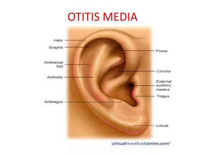
ANATOMY OF THE EAR
What Causes Otitis Media
Otitis media • Otitis media (Latin for "inflammation of the middle ear") is the medical term for middle ear infection.
AETIOLOY • The common cause of all forms of otitis media is blockage of the Eustachian tube. This is usually due to swelling of the mucous membranes in the nasopharynx, which in turn can be caused by a viral upper respiratory infection or by allergies.[2] This is seen as a progression from a Type A tympanogram to a Type C to a Type B tympanogram. • ]
PATHOGENESIS • Because of the blockage of the Eustachian tube, the air volume in the middle ear is trapped and parts of it are slowly absorbed by the surrounding tissues, leading to a mild vacuum in the middle ear. Eventually the vacuum can reach a point where fluid from the surrounding tissues is sucked in to the middle ear's cavity (also called tympanic cavity), causing middle ear effusion.
By reflux or suction of material from the nasopharynx into the normally sterile middle ear space, the fluid may then become infected - usually with bacteria. In rare cases, however, the virus that caused the initial upper respiratory tract infection can itself be identified as the pathogen causing the infection in the middle ear.[2
Signs and symptoms • An integral symptom of acute otitis media is ear pain; other possible symptoms include fever, and irritability (in infants). Since an acute otitis media is usually precipitated by an upper respiratory tract infection, there often are accompanying symptoms like cough and nasal blockage& discharge.[1]
Diagnosis • As its typical symptoms overlap with other conditions, clinical history alone is not sufficient to predict whether acute otitis media is present; it has to be complemented by visualization of thetympanic membrane.[3]
To confirm the diagnosis, middle ear effusion and inflammation of the eardrum have to be identified; signs of these are fullness, bulging, cloudiness and redness of the eardrum.[1] Viral otitis may also result in blisters on the external side of the tympanic membrane, which is called bullousmyringitis (myringa being Latin for "eardrum")[4]
fullness, bulging, cloudiness and redness of the eardrum
However, sometimes even examination of the eardrum may not be able to confirm the diagnosis, especially if the canal is small and there is wax in the ear that obscures a clear view of the eardrum. Also, an upset child's crying can cause the eardrum to look inflamed due to distension of the small blood vessels on it, mimicking the redness associated with otitis media.
TYPES OF OTITIS MEDIA • Acute otitismedia(AOM) • Otitis media with effusion(OME) • Chronic suppurativeotitismedia(CSOM) • Adhesive otitismedia(Adh OM)
Acute otitis media • Acute otitis media (AOM) is usually developing on the basis of a (viral) upper respiratory infection with blockage of the Eustachian tube and effusion in the middle ear, when the fluid in the middle ear gets additionally infected with bacteria. The most common bacteria found in this case are Streptococcus pneumoniae, Haemophilusinfluenzae, and Moraxellacatarrhalis.[1]
Otitis media with effusion • Otitismedia with effusion (OME), also called serous or secretoryotitis media (SOM) or glue ear,[5] is simply a collection of fluid that occurs within the middle ear space due to the negative pressure produced by altered Eustachian tube function.
This can occur purely from a viral URI, with no pain or bacterial infection, or it can precede and/or follow acute bacterial otitis media. Fluid in the middle ear sometimes causes conductive hearing impairment, but only when it interferes with the normal vibration of the eardrum by sound waves.
Over weeks and months, middle ear fluid can become very thick and glue-like (thus the name glue ear), which increases the likelihood of its causing conductive hearing impairment. Early-onset OME is associated with feeding while lying down and early entry into group child care, parental smoking, too short a period of breastfeeding and greater amounts of time spent in group child care increased the duration of OME in the first two years of life.[6]
Chronic suppurativeotitis media • Chronic suppurativeotitis media involves a perforation (hole) in the tympanic membrane and active bacterial infection within the middle ear space for several weeks or more
. There may be enough pus that it drains to the outside of the ear (otorrhea), or the purulence may be minimal enough to only be seen on examination using a binocular microscope. This disease is much more common in persons with poor Eustachian tube function. Hearing impairment often accompanies this disease.
Adhesive otitis media • Adhesive otitis media has occurred when a thin retracted ear drum becomes sucked into the middle ear space and stuck, i.e. adherent, to the ossicles and other bones of the middle ear.
Diagnosis To differantiate between the typesofotitismediais , by visualization of thetympanic membrane.[3] • History • Examination - Otoscopic - Microscopic
Prevention • Long term antibiotics, while they decrease rates of infection during treatment, have an unknown effect on long term outcomes such as hearing loss.[7] They are thus not recommended.[1]
Pneumococcal conjugate vaccines when given during infancy decrease rates of acute otitis media by 6–7% and if implemented broadly would have a significant public health benefit.[8]
Certain factors such as season, allergy predisposition and of older siblings are known to be determipresencenantsof recurrent otitis media and persistent middle ear effusions (MEE).[9]Previous history of recurrence, environmental exposure to tobacco smoke, use of daycare, and lack of breastfeeding have all been associated with increased risk of OM development, recurrence, and persistent MEE.[10][11]
There is some evidence that breastfeeding for the first twelve months of life is associated with a reduction in the number and duration of OM infections.[12][13] Pacifier use, on the other hand, has been associated with more frequent episodes of AOM.[14]
Evidence does not support zinc supplementation as an effort to reduce otitis rates except maybe in those with severe malnutrition such as marasmus.[15]
Prognosis Complications of acute otitis media consist in perforation of the ear drum, infection of the mastoid space behind the ear, or bacterial meningitis in rare cases.[29][30]
Rupture • In severe or untreated cases, the tympanic membrane may rupture, allowing the pus in the middle ear space to drain into the ear canal. If there is enough of it, this drainage may be obvious. Even though the rupture of the tympanic membrane suggests a highly painful and traumatic process, it is almost always associated with the dramatic relief of pressure and pain. In a simple case of acute otitis media in an otherwise healthy person, the body's defenses are likely to resolve the infection and the ear drum nearly always heals. • Hearing loss • Children with recurrent episodes of acute otitis media and those with otitis media with effusion or chronic otitis media, have higher risks of developing conductive and sensorineural hearing loss. Globally approximately 141 million people have mild hearing loss due to otitis media (2.1% of the population).[31] This is more common in males (2.3%) than females (1.8%).[31]
Management • Oral and topical analgesics are effective to treat the pain caused by otitis media. Oral agents include ibuprofen, paracetamol (acetaminophen), and opiates. Topical agents shown to be effective include antipyrine and benzocaine ear drops.[16]Decongestants and antihistamines, either nasal or oral, are not recommended due to the lack of benefit and concerns regarding side effects.[17]
References • ^ Jump up to: abcdefgLieberthal, AS; Carroll, AE; Chonmaitree, T; Ganiats, TG; Hoberman, A; Jackson, MA; Joffe, MD; Miller, DT; Rosenfeld, RM; Sevilla, XD; Schwartz, RH; Thomas, PA; Tunkel, DE (2013 Feb 25). "The Diagnosis and Management of Acute Otitis Media.". Pediatrics131 (3): e964–99. doi:10.1542/peds.2012-3488. PMID23439909. • ^ Jump up to: ab John D Donaldson. "Acute Otitis Media". Medscape. Retrieved 17 March 2013. • Jump up ^Laine MK, Tähtinen PA, Ruuskanen O, Huovinen P, Ruohola A (May 2010). "Symptoms or symptom-based scores cannot predict acute otitis media at otitis-prone age". Pediatrics125 (5): e1154–61. doi:10.1542/peds.2009-2689. PMID20368317. • Jump up ^ Roberts DB (April 1980). "The etiology of bullousmyringitis and the role of mycoplasmas in ear disease: a review". Pediatrics65 (4): 761–6. PMID7367083. • Jump up ^"Glue Ear". NHS Choices. Department of Health. Retrieved 3 November 2012. • Jump up ^ Owen MJ, Baldwin CD, Swank PR, Pannu AK, Johnson DL, Howie VM (1993). "Relation of infant feeding practices, cigarette smoke exposure, and group child care to the onset and duration of otitis media with effusion in the first two years of life". J. Pediatr.123 (5): 702–11. doi:10.1016/S0022-3476(05)80843-1. PMID8229477. • Jump up ^ Leach AJ, Morris PS (2006). "Antibiotics for the prevention of acute and chronic suppurativeotitis media in children". In Leach, Amanda J. Cochrane Database Syst Rev (4): CD004401. doi:10.1002/14651858.CD004401.pub2. PMID17054203. • Jump up ^ Jansen AG, Hak E, Veenhoven RH, Damoiseaux RA, Schilder AG, Sanders EA (2009). "Pneumococcal conjugate vaccines for preventing otitis media". In Jansen, Angelique GSC. Cochrane Database Syst Rev (2): CD001480. doi:10.1002/14651858.CD001480.pub3. PMID19370566. • Jump up ^ Rovers MM, Schilder AG, Zielhuis GA, Rosenfeld RM (2004). "Otitis media". Lancet363: 564–573. doi:10.1016/S0140-6736(04)15546-3. PMID14962529. • Jump up ^Pukander J, Luotonem J, Timonen M, Karma P (1985). "Risk factors affecting the occurrence of acute otitis media among 2-3 year old urban children". ActaOtolaryngol100 (3-4): 260–265. PMID4061076. • Jump up ^Etzel RA (1987). "Smoke and ear effusions". Pediatrics79 (2): 309–311. PMID3808812. • Jump up ^ Dewey KG, Heinig MJ, Nommsen-Rivers LA (1995). "Differences in morbidity between breast-fed and formula-fed infants". J Pediatr126 (5 Pt 1): 696–702. doi:10.1016/S0022-3476(95)70395-0. PMID7751991. • Jump up ^ Saarinen UM (1982). "Prolonged breast feeding as prophylaxis for recurrent otitis media". ActaPediatr Scan71 (4): 567–571. PMID7136672. • Jump up ^ Rovers MM, Numans ME, Langenbach E, Grobbee DE, Verheij TJ, Schilder AG (August 2008). "Is pacifier use a risk factor for acute otitis media? A dynamic cohort study". FamPract25 (4): 233–6. doi:10.1093/fampra/cmn030. PMID18562333. • Jump up ^ Abba K, Gulani A, Sachdev HS (2010). "Zinc supplements for preventing otitis media". In Abba, Katharine. Cochrane Database Syst Rev (2): CD006639. doi:10.1002/14651858.CD006639.pub2. PMID20166086. • Jump up ^Sattout, A.; Jenner, R. (February 2008). "Best evidence topic reports. Bet 1. The role of topical analgesia in acute otitis media". Emerg Med J25 (2): 103–4. doi:10.1136/emj.2007.056648. PMID18212148. • Jump up ^ Coleman C, Moore M (2008). "Decongestants and antihistamines for acute otitis media in children". In Coleman, Cassie. Cochrane Database Syst Rev (3): CD001727. Study 2010.". Lancet380 (9859): 2095–128. doi:10.1016/S0140-6736(12)61728-0. PMID23245604. • External links
doi:10.1002/14651858.CD001727.pub4. PMID18646076. • Jump up ^Venekamp, Roderick P (31 January 2013). "Antibiotics for acute otitis media in children (Systematic Review)". Cochrane Database of Systematic Reviews (1): CD000219 (3rd rev.). doi:10.1002/14651858.CD000219.pub3. PMID14973951. • Jump up ^Damoiseaux R (2005). "Antibiotic treatment for acute otitis media: time to think again". CMAJ172 (5): 657–8. doi:10.1503/cmaj.050078. PMC550637. PMID15738492. • Jump up ^Marchetti F, Ronfani L, Nibali S, Tamburlini G (2005). "Delayed prescription may reduce the use of antibiotics for acute otitis media: a prospective observational study in primary care". Arch PediatrAdolesc Med159 (7): 679–84. doi:10.1001/archpedi.159.7.679. PMID15997003. • Jump up ^Kozyrskyj A, Klassen TP, Moffatt M, Harvey K (2010). "Short-course antibiotics for acute otitis media". Cochrane Database of Systematic Reviews (9): CD001095 (2nd rev.). doi:10.1002/14651858.CD001095.pub2. PMID20824827. • Jump up ^Gulani A, Sachdev HP, Qazi SA (January 2010). "Efficacy of short course (<4 days) of antibiotics for treatment of acute otitis media in children: a systematic review of randomized controlled trials". Indian Pediatr47 (1): 74–87. doi:10.1007/s13312-010-0010-9. PMID19736367. • Jump up ^ McDonald S, Langton Hewer CD, Nunez DA (2008). "Grommets (ventilation tubes) for recurrent acute otitis media in children". In McDonald, Stephen. Cochrane Database Syst Rev (4): CD004741. doi:10.1002/14651858.CD004741.pub2. PMID18843668. • Jump up ^ Browning GG, Rovers MM, Williamson I, Lous J, Burton MJ (2010). "Grommets (ventilation tubes) for hearing loss associated with otitis media with effusion in children". In Browning, George G. Cochrane Database Syst Rev (10): CD001801. doi:10.1002/14651858.CD001801.pub3. PMID20927726. • Jump up ^ Wilson SA, Mayo H, Fisher M (May 2003). "Clinical inquiries. Are tympanostomy tubes indicated for recurrent acute otitis media?" (PDF). J FamPract52 (5): 403–4, 406. PMID12737775. • Jump up ^ Rosenfeld RM, Culpepper L, Yawn B, Mahoney MC (June 2004). "Otitis media with effusion clinical practice guideline". Am Fam Physician69 (12): 2776, 2778–9. PMID15222643. • Jump up ^ Pratt-Harrington D (October 2000). "Galbreath technique: a manipulative treatment for otitis media revisited". J Am Osteopath Assoc100 (10): 635–9. PMID11105452. • Jump up ^Bronfort G, Haas M, Evans R, Leininger B, Triano J (2010). "Effectiveness of manual therapies: the UK evidence report". ChiroprOsteopat18: 3. doi:10.1186/1746-1340-18-3. PMC2841070. PMID20184717. • Jump up ^Spremo S, Udovcić B (2007 May). "Acute mastoiditis in children: susceptibility factors and management". Bosn J Basic Med Sci.7 (2): 127–31. PMID17489747. • Jump up ^Klossek JM (2009 Jul-Aug). "Recherche et prise en charge de la ported'entrée ORL des méningitesaiguësbactériennescommunautaires" [Diagnosis and management of ENT conditions responsible for acute community acquired bacterial meningitis]. Médecine et Maladies Infectieuses (in French) 39 (7–8): 554–9. doi:10.1016/j.medmal.2009.02.027. PMID19419828. • ^ Jump up to: abVos, T (2012 Dec 15). "Years lived with disability (YLDs) for 1160 sequelae of 289 diseases and injuries 1990-2010: a systematic analysis for the Global Burden of Disease Study 2010.". Lancet380 (9859): 2163–96. doi:10.1016/S0140-6736(12)61729-2. PMID23245607. • Jump up ^Da Costa SS; Rosito, Letícia Petersen Schmidt; Dornelles, Cristina (February 2009). "Sensorineural hearing loss in patients with chronic otitis media". Eur Arch Otorhinolaryngol266 (2): 221–4. doi:10.1007/s00405-008-0739-0. PMID18629531. • Jump up ^ Roberts K (June 1997). "A preliminary account of the effect of otitis media on 15-month-olds' categorization and some implications for early language learning". J Speech Lang Hear Res40 (3): 508–18. PMID9210110. • Jump up ^Bidadi S, Nejadkazem M, Naderpour M (November 2008). "The relationship between chronic otitis media-induced hearing loss and the acquisition of social skills". Otolaryngol Head Neck Surg139 (5): 665–70. doi:10.1016/j.otohns.2008.08.004. PMID18984261. • Jump up ^Gouma P, Mallis A, Daniilidis V, Gouveris H, Armenakis N, Naxakis S (January 2011). "Behavioral trends in young children with conductive hearing loss: a case-control study". Eur Arch Otorhinolaryngol268 (1): 63–6. doi:10.1007/s00405-010-1346-4. PMID20665042. • Jump up ^Yilmaz S, Karasalihoglu AR, Tas A, Yagiz R, Tas M (February 2006). "Otoacoustic emissions in young adults with a history of otitis media". J LaryngolOtol120 (2): 103–7. doi:10.1017/S0022215105004871. PMID16359151. • Jump up ^ John D Donaldson. "Acute Otitis Media". Medscape. Retrieved 17 March 2013. • Jump up ^ Lozano, R (2012 Dec 15). "Global and regional mortality from 235 causes of death for 20 age groups in 1990 and 2010: a systematic analysis for the Global Burden of Disease
- More by User

otitis media
Definition. Inflammation of the middle ear cleftTypesAcute viral Otitis MediaAcute Bacterial Otitis MediaAcute Necrotising Otitis MediaOtitis Media with EffusionTuberculous Otitis MediaChronic Suppurative Otitis Media. Acute Otitis Media. Acute infection of middle ear cleft with presence of middle ear effusion and signs of middle ear inflammation: AAP Recurrent otitis media is defined as 3 or more episodes in 6 months or 4 or more in a year .
1.47k views • 36 slides

Otitis Media
1.04k views • 41 slides

OTITIS MEDIA. By: Anitha Jacob PA-S November 8, 2000. OTITIS MEDIA. Definition: Presence of a middle ear infection Acute Otitis Media: occurrence of bacterial infection within the middle ear cavity.
1.02k views • 18 slides

Otitis Media. Dr John Curotta Head of ENT Surgery The Children’s Hospital at Westmead. What is Otitis Media?. AOM = Acute OM OME = OM with Effusion (= ‘glue ear’) CSOM = Chronic Suppurative Otitis Media ( = a hole in the ear drum
994 views • 50 slides

Otitis media
Otitis media. 2 ½ year old girl Generally well. Attends nursery school Recent course of Augmentin (2 weeks prior) Mon 13 September 2010 – bilateral conjunctivitis; no fever, otherwise well; mild discharge; no preauricular lymph nodes After 3 days gave Tobrex eye drops.
867 views • 48 slides

Otitis Media. Slide presentation from; Kevin Katzenmeyer , MD Ronald W. Deskin , MD Dept. of Oto -HNS UTMB-Galveston February 17, 1999. Otitis Media - Definition. Inflammation of the middle ear May also involve inflammation of mastoid, petrous apex, and perilabyrinthine air cells.
348 views • 14 slides

OTITIS MEDIA . Our Ear . What is otitis media?. Otitis media Latin for "Middle otitis " It is one of the two categories of ear inflammation that can underlie what is commonly called an earache , the other being otitis externa . . Classifications of Otitis Media .
3.43k views • 17 slides

Otitis media. Terminology. Otitis Media: inflammation of the middle ear cleft or mucosa. Acute Less than 6 weeks Chronic More than 6 weeks Recurrent acute otitis media 3 episodes/6 months or 4 or more episodes/1 year
937 views • 24 slides

OTITIS MEDIA. Babak Saedi Imam Khomeini Hospital. OTITIS MEDIA. Definition: Presence of a middle ear infection Acute Otitis Media: occurrence of bacterial infection within the middle ear cavity.
664 views • 22 slides

Otitis Media. Mary Bennett, Amanda Buisman & Roline Campbell. Pertinent Anatomy. Ossicles (malleus, incus, stapes). OR Auricle. External Ear Canal. OR Tympanic Membrane. Pertinent Anatomy. (Cone of light). Physiology of the Ear. External Ear
2.36k views • 86 slides

Acute Otitis Media
Acute Otitis Media. Objectives. Define otitis media (OM), acute otitis media (AOM) and otitis media with effusion (OME) Be familiar with the epidemiology of AOM List causative pathogens in children with AOM and current bacteriologic resistance patterns .
824 views • 24 slides

Otitis Media. 5 y/o Female Incomplete cleft of secondary palate Pain in left ear Tubes 4 years ago. No Medications Cleft has been repaired in 2001 and has healed well. OM Case 1. Otoscopy. Right ear canal clear TM intact Amber colored fluid behind left TM. Tymps. SRT/WR. Audio.
725 views • 51 slides

OTITIS MEDIA. The most important disease of the middle ear and mastoid are inflammations of various kinds and hearing loses. Tumors of the middle ear are rare . In this chapter we'll mainly discussed acute suppurative otitis media (ASOM) and chronic suppurative otitis media (CSOM). ASOM.
1.67k views • 44 slides

Otitis Media. Otitis Media. Most common reason for visit to pediatrician Tympanostomy tube placement is 2nd most common surgical procedure in children Development of multidrug-resistant bacteria. Otitis Media - Definition. Inflammation of the middle ear
580 views • 29 slides

Otitis Media. K. Myra Lalas Peds PGY 2. Definitions. 1. Otitis media with effusion (OME)- presence of middle ear effusion without sings or symptoms of infection
1.31k views • 26 slides
Chronic otitis media
Chronic otitis media. Chunfu Dai M.D & Ph. D Otolaryngology Department Eye Ear Nose and Throat Hospital Fudan University. Definition. COM: unresolved inflammatory process of the middle ear and mastoid associated with TM perforation, otorrhea and hearing loss. Etiology.
452 views • 25 slides

IMAGES
VIDEO
COMMENTS
6. 6 | P a g e Otitis media with effusion (OME), also known as serous otitis media (SOM) or secretory otitis media (SOM), and colloquially referred to as 'glue ear,'[25] is fluid accumulation that can occur in the middle ear and mastoid air cells due to negative pressure produced by dysfunction of the Eustachian tube. This can be associated with a viral URI or bacterial infection such as ...
10 likes • 12,142 views. M. Makbul Hussain Chowdhury. It is a chronic inflammation of the middle ear and the mastoid cavity. Health & Medicine. 1 of 17. Download Now. Download to read offline. CASE PRESENTATION ON CHRONIC SUPPURATIVE OTITIS MEDIA - Download as a PDF or view online for free.
Acute otitis media is extremely common in children. In fact, it is one of the most common diagnosis in children who are seen in outpatient settings, and is one of the most common reasons for antibiotic therapy. The peak incidence of AOM is between 6 months and 2 years of age. Three out of four children will experience at least one ear infection ...
Otitis media (OM) with effusion (OME) often follows an episode of AOM. Consider OME in patients with recent AOM in whom the history includes any of the following symptoms: Hearing loss - Most young children cannot provide an accurate history; parents, caregivers, or teachers may suspect a hearing loss or describe the child as inattentive.
1 A 2 year old boy with Acute Otitis Media - Case Presentation. 2 Nilanjana Basu, Homoeopathic Physician Sameer Rana E.N.T. Specialist, Lecturer, Department of ... Download ppt "A 2 year old boy with Acute Otitis Media - Case Presentation" Similar presentations . Otitis Media Lawrence Pike. Chapter 6 Fever Case I.
Read chapter 2 of Infectious Diseases: A Case Study Approach online now, exclusively on AccessPharmacy. AccessPharmacy is a subscription-based resource from McGraw Hill that features trusted pharmacy content from the best minds in the field.
The history of acute otitis media (AOM) varies with age, but a number of constant features manifest during the otitis-prone years. In the neonate, irritability or feeding difficulties may be the only indication of a septic focus. Older children begin to demonstrate a consistent presence of fever (with or without a coexistent upper respiratory ...
What is Otitis Media". Otitis Media" means inflammation of the middle earDifferent types:Acute: presence of fluid, pus, redness of eardrum and possible feverChronic: fluid lasting 6 weeks or moreMay or may not be infectedDifferent types = different treatmentTypically when a physician says ear infection, actually means acute otitis media".
47 Brain Abscess. Download ppt "ACUTE AND CHRONIC OTITIS MEDIA". Objectives To define acute otitis media (AOM) and chronic otitis media (COM) To understand the clinical presentation and diagnostic evaluation of AOM and COM To define the various types of cholesteatoma and how they develop. To provide an overview of the management of AOM and COM.
CASE PRESENTATION- ACUTE OTITS MEDIA - Free download as Powerpoint Presentation (.ppt / .pptx), PDF File (.pdf), Text File (.txt) or view presentation slides online.
a.Atico-antral chronic otitis. a.Serous Otitis media- Stages: URTI. hearing loss . Medical management- careful. Assessment:- Collect health. Pain R/T. Disturbed sensory. Otitis media - Download as a PDF or view online for free.
Case report acute otitis media - Free download as Powerpoint Presentation (.ppt / .pptx), PDF File (.pdf), Text File (.txt) or view presentation slides online. overview of OMA
Chronic suppurative otitis media (CSOM) is a perforated tympanic membrane with persistent drainage from the middle ear (ie, lasting >6-12 wk). Chronic suppuration can occur with or without cholesteatoma, and the clinical history of both conditions can be very similar. ... Regardless of the presentation, imaging to define the abscess should be ...
PPT Otitis Media - Free download as Powerpoint Presentation (.ppt / .pptx), PDF File (.pdf), Text File (.txt) or view presentation slides online. This case study aims to identify and determine the health problem and needs of the patient who underwent for Otitis Media with effusion. It aims to give an effective nursing care plan to the patient 3.
Definition: long-standing infection of a part or whole of the middle ear cleft characterized by ear discharge and a permanent perforation. Classification: anatomical or pathological. Etiology: Ascending infection, allergy and ASOM. Diagnosis: history, examination and investigations. History: Ear discharge and hearing loss.
Acute otitis media is defined as an infection of the middle ear space. It is a spectrum of diseases that includes acute otitis media (AOM), chronic suppurative otitis media (CSOM), and otitis media with effusion (OME). Acute otitis media is the second most common pediatric diagnosis in the emergency department, following upper respiratory infections. Although otitis media can occur at any age ...
Presentation Transcript. Acute Otitis Media Dr. Ghaleb Zughayar Consultant Pediatrician and Neonatologist. Definition • Acute Otitis Media (AOM) • "acute onset of symptoms, evidence of a middle ear effusion, and signs or symptoms of middle ear inflammation.". • Otitis Media with effusion (OME) • "Presence of MEE without signs or ...
Presentation Transcript. Acute Otitis Media Dr. Hamid Rahimi Pediatric Infectious Disease Specialist. Acute Otitis Media • The most common infection for which antibacterial agents are prescribed for children in the US • 1/3 of office visits to pediatricians • Peak incidence 6 - 12 months old • ≈ 2/3 of children experience at least ...
Otitis media. Dec 5, 2019 •. 55 likes • 16,008 views. Abhay Rajpoot. Otitis media is a group of inflammatory diseases of the middle ear. The two main types are acute otitis media (AOM) and otitis media with effusion (OME). AOM is an infection of rapid onset that usually presents with ear pain. Read more.
Acute Otitis Media. By Jennifer Naruta, RN. What is Otitis Media?. An acute infection of the middle ear Often follows eustachian tube dysfunction (ETD) or URI. Incidence of OM…. Most episodes occur in first 24 months of life 50% of infants in U.S. have first AOM before 6 months. Download Presentation. doses.
Classifications of Otitis Media • Chronic suppurativeotitismedia involves a perforation in the eardrum and active bacterial infection within the middle ear space for several weeks or more. There may be enough pus that it drains to the outside of the ear. Hearing impairment often accompanies this disease. Acute otitis media.
Adhesive otitis media • Adhesive otitis media has occurred when a thin retracted ear drum becomes sucked into the middle ear space and stuck, i.e. adherent, to the ossicles and other bones of the middle ear. Diagnosis To differantiate between the typesofotitismediais , by visualization of thetympanic membrane. [3] •.